Understanding MRI Scans: What They Show
DS
By Deepak Sharma, DC
Medically reviewed
Owner & Doctor of Chiropractic · 30 min read · Published · Updated · Last reviewed
Introduction
Recovering from an accident or dealing with an injury can be a confusing and painful process. Getting a clear picture of the internal damage is crucial for accurate diagnosis and effective treatment. While X-rays and CT scans provide valuable information, they have limitations, particularly when it comes to visualizing soft tissues like muscles, ligaments, tendons, and nerves. This is where Magnetic Resonance Imaging, commonly known as an MRI scan, plays a critical role. For residents of Beaumont, TX, understanding what an MRI scan Beaumont TX can reveal is essential, especially after experiencing trauma or persistent pain.
An MRI scan is a non-invasive diagnostic tool that uses powerful magnetic fields and radio waves to create detailed cross-sectional images of the body. Unlike X-rays or CT scans, it does not use ionizing radiation. This makes it a safer option for many patients, particularly those requiring multiple scans. For accident victims and those suffering from injuries in the Beaumont area, access to reliable and detailed diagnostic imaging is paramount. At Car Accident Cares, we understand the urgency and the need for clarity. We are committed to providing accessible diagnostic imaging Beaumont, including MRI scans Beaumont TX, with the added benefit of 24/7 availability and transparent, affordable pricing to support your journey towards recovery.
What Is an MRI Scan?
To truly appreciate the value of an MRI scan Beaumont TX in diagnosing injuries, it helps to understand the fundamental nature of this advanced imaging technology. This section provides foundational knowledge about what an MRI is and how it differs from other imaging methods.
Defining Magnetic Resonance Imaging
MRI stands for Magnetic Resonance Imaging. Breaking down the name helps explain the process:
- Magnetic: The technology uses a strong magnetic field.
- Resonance: Radio waves are sent into the body, causing hydrogen protons which are abundant in water and fat within tissues to “resonate” or align with the magnetic field.
- Imaging: When the radio waves are turned off, the protons release energy signals. These signals are detected by receiver coils and processed by a computer to create detailed images of internal body structures.
Unlike X-rays and CT scans, which primarily visualize bone and dense tissues, MRI excels at visualizing soft tissues. This is particularly important in cases of injuries resulting from car accidents or other trauma, where damage to muscles, ligaments, tendons, and nerves is common but may not be clearly visible on other imaging modalities. This superior soft tissue contrast is a key reason why an MRI scan Beaumont TX is often recommended for evaluating these types of injuries.
Key Components of MRI Technology
An MRI machine is a complex piece of equipment, but its key components include:
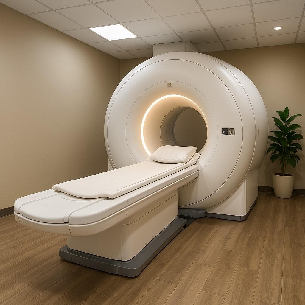
- Superconducting Magnet: This is the largest and most recognizable part of the MRI scanner, typically shaped like a large tube or a donut. It generates a very strong, stable magnetic field, often measured in Teslas (T). Common field strengths are 1.5T and 3T. Higher field strengths generally produce higher-resolution images.
- Gradient Coils: These smaller magnets within the main magnet create varying magnetic fields across the body. These gradients allow the scanner to pinpoint the location of the signals coming from the hydrogen protons, enabling the creation of cross-sectional images.
- Radiofrequency (RF) Transmitter/Receiver: This component sends radio waves into the body and receives the signals emitted by the protons after the RF pulse is turned off. The receiver coils are designed to be placed near the body part being scanned to capture these signals effectively.
MRI systems come in different designs. Closed MRI systems are the traditional tube shape, which can sometimes be challenging for patients with claustrophobia. Open MRI systems have a less enclosed design, offering a more comfortable experience for some individuals, though they may use lower magnetic field strengths affecting image quality in some cases. When seeking an MRI scan Beaumont TX, understanding the type of system available can be helpful.
What Makes MRI Unique Among Imaging Modalities
Several features distinguish MRI from other diagnostic imaging techniques, making it uniquely valuable for certain types of injuries and conditions:
- No Ionizing Radiation: This is a major advantage, especially for patients who may require multiple scans or for sensitive populations like children or pregnant women. Unlike X-rays or CT scans, there is no cumulative radiation exposure risk with MRI.
- Superior Soft Tissue Contrast: MRI provides unparalleled detail in visualizing soft tissues like muscles, ligaments, tendons, cartilage, nerves, and the spinal cord. This is crucial for diagnosing injuries like muscle tears, ligament sprains including whiplash, and herniated discs – common issues after car accidents in the Beaumont area.
Multi-Planar Imaging: MRI can generate images in multiple planes (axial, sagittal, coronal) without repositioning the patient. This allows for a comprehensive view of the injured area from different angles, aiding in accurate diagnosis.
These unique capabilities make MRI scans Beaumont TX an indispensable tool in the diagnostic arsenal, particularly for evaluating complex injuries that may not be clearly visible on other scans. To understand how MRI compares to other imaging options available, explore our dedicated Diagnostic Imaging service.
How MRI Scans Work: The Science Behind the Images
While you don’t need a degree in physics to get an MRI scan Beaumont TX, understanding the basic science behind it can demystify the process and highlight its precision. The images produced by MRI are a result of manipulating the behavior of hydrogen protons within your body’s tissues using magnetic fields and radio waves.
The Physics of Magnetic Resonance
The human body is largely composed of water molecules, which contain hydrogen atoms. Each hydrogen atom has a single proton at its nucleus. These protons behave like tiny magnets, spinning and normally pointing in random directions.
When placed in the strong magnetic field of the MRI scanner, these hydrogen protons align with the direction of the magnetic field, much like compass needles aligning with the Earth’s magnetic field. The stronger the MRI magnet (e.g., 3T vs. 1.5T), the better the alignment.
Next, a radiofrequency (RF) pulse is applied to the body. This pulse is specifically tuned to the frequency at which the aligned protons are “resonating” (spinning) in that particular magnetic field strength. The RF pulse temporarily knocks the protons out of alignment.
When the RF pulse is turned off, the protons realign with the main magnetic field. As they realign, they release the absorbed energy in the form of weak radio signals. Different tissues like bone, fat, muscle, and water have different compositions and molecular environments, causing their hydrogen protons to realign at slightly different rates and emit signals of varying strengths.
The MRI scanner’s receiver coils detect these subtle differences in signals. These signals are then sent to a powerful computer, which processes the data and uses complex mathematical algorithms to construct detailed cross-sectional images. Think of it like receiving “cellular radio signals” from inside your body, with different tissues sending back unique patterns that the computer can translate into a visual representation.
Safety Considerations in Beaumont Scans
MRI is generally considered a very safe imaging modality due to the absence of ionizing radiation. However, there are important safety considerations related to the strong magnetic field. When preparing for an MRI scan Beaumont TX, you will undergo a thorough screening process.
- Magnetic Field Strength: The magnet is always on, even when the machine is not scanning. Metallic objects can be strongly attracted to the magnet, posing a safety risk. This is why it’s crucial to remove all metal items jewelry, watches, coins, etc. before entering the MRI suite.
- Medical Implants: Certain medical implants can be affected by the magnetic field or the radio waves. It is absolutely critical to inform your healthcare provider and the MRI technologist about any implants you have, including pacemakers, defibrillators, cochlear implants, certain types of clips used during brain surgery, and some metal plates or screws. Some implants are “MRI-safe,” while others are “MRI-conditional” safe under specific conditions, and some are “MRI-unsafe.” Thorough screening ensures your safety.
- Claustrophobia: Being in an enclosed space can be anxiety-inducing for some individuals. If you experience claustrophobia, discuss this with your doctor or the MRI facility beforehand. Options like open MRI systems if available, medication to reduce anxiety, or using relaxation techniques can help make the experience more comfortable. Our facility in Beaumont aims to address these concerns to ensure a smooth process.
- Noise: The MRI machine makes loud knocking or buzzing noises during the scan, caused by the gradient coils rapidly turning on and off. You will be provided with earplugs or headphones to protect your hearing and improve comfort.
A strict screening protocol is in place at our MRI scan Beaumont TX facility to ensure your safety. You will be asked to fill out a detailed questionnaire about any metal in or on your body. Honesty and thoroughness in this screening are paramount.
Advanced MRI Sequences and Their Uses
Modern MRI scanners utilize various sequences, which are different ways of applying magnetic fields and radio pulses, to highlight different tissue characteristics and visualize specific types of abnormalities. Understanding these can help appreciate the depth of information an MRI scan Beaumont TX provides.
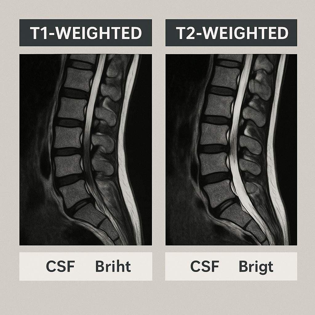
- T1-weighted images: These images are good for visualizing anatomical detail. Fat appears bright, while water and fluid like cerebrospinal fluid, CSF appear dark. They are often used to assess the anatomy of the brain, spinal cord, and other structures.
- T2-weighted images: These images are excellent for highlighting areas of inflammation, edema swelling, or fluid collection. Water and fluid appear bright, while fat appears gray-ish, and most solid tissues are darker than fluid. They are often used to detect injuries, tumors, or inflammation.
- FLAIR (Fluid Attenuated Inversion Recovery): This is a type of T2-weighted sequence where the signal from CSF is suppressed. It is particularly useful for visualizing lesions in the brain that are close to CSF spaces, such as those caused by multiple sclerosis or certain types of brain injuries.
- DWI (Diffusion-Weighted Imaging): This sequence measures the random motion (diffusion) of water molecules in tissues. It is highly sensitive to early stroke, certain tumors, and some types of brain injury, including diffuse axonal injury (DAI) which can occur in traumatic brain injuries.
- Contrast-Enhanced Protocols: In some cases, a contrast agent typically Gadolinium-based is injected into a vein before the scan. This agent changes the magnetic properties of tissues, causing them to appear brighter on the images. Contrast is often used to enhance the visibility of tumors, inflammation, or blood vessels.
Note: There are ACR guidelines regarding contrast use, especially in certain populations like pregnant women.
By using a combination of these sequences, radiologists can gain a comprehensive understanding of the tissues being imaged and accurately diagnose a wide range of conditions and injuries.
Injuries and Conditions Diagnosed by MRI in Beaumont
The ability of MRI to provide detailed images of soft tissues makes it an invaluable diagnostic tool for a wide array of injuries and conditions, particularly those resulting from trauma like car accidents common in the Beaumont area. For anyone seeking an MRI scan Beaumont TX after an injury, knowing what the scan can reveal is reassuring.
Soft Tissue Trauma from Car Accidents
Car accidents are a frequent cause of injury in Beaumont, and while bones might be checked with X-rays, soft tissue injuries often require MRI for proper diagnosis. An MRI scan Beaumont TX can clearly visualize:
- Muscle Tears and Strains: Shows the location and extent of tears within muscle fibers.
- Ligament Sprains: Visualizes tears or stretching of ligaments, such as those in the neck (whiplash) or back. Whiplash-associated disorders involve injuries to ligaments, muscles, and discs in the neck, and MRI is crucial for assessing the severity. See how MRI guides whiplash neck pain treatment.
- Tendon Injuries: Detects tears or inflammation in tendons that connect muscles to bones.
These soft tissue injuries are often the primary source of pain and dysfunction after a crash, and accurate identification via MRI is essential for guiding appropriate therapy and rehabilitation.
Spinal Injuries Revealed by MRI
The spine is particularly vulnerable in accidents. MRI scan Beaumont TX provides detailed images of the spinal cord, nerve roots, discs, ligaments, and facet joints, allowing for the diagnosis of critical spinal injuries:
- Herniated or Bulging Discs: MRI is the gold standard for visualizing intervertebral discs and determining if they are bulging or herniated and pressing on nerve roots or the spinal cord. This information is vital for planning treatment, whether it’s physical therapy, injections, or surgery. Get an MRI-based disc injury diagnosis by consulting a Herniated Disc Doctor.
- Spinal Cord Compression: Clearly shows if the spinal cord is being compressed by disc material, bone spurs, or other masses.
- Facet Joint Damage: Visualizes inflammation or degenerative changes in the small joints on the back of the spine that can be a significant source of chronic back pain.
- Ligamentous Injuries of the Spine: Detects tears or instability in the strong ligaments that support the spinal column, which can occur in severe trauma.
Given the unfortunate frequency of auto accidents, having access to precise diagnostic imaging Beaumont like MRI is critical for assessing potential spinal injuries.
Traumatic Brain Injuries (TBI) Detection
Following a head injury, especially in a car accident, a CT scan is often the first imaging test performed because it is quick and excellent at detecting acute bleeding and skull fractures. However, CT can miss certain types of brain injuries, particularly those affecting the white matter, like Diffuse Axonal Injury (DAI).
An MRI scan Beaumont TX, especially utilizing sequences like DWI and FLAIR, is much more sensitive than CT in detecting DAI, which involves microscopic tearing of nerve fibers throughout the brain. MRI can also better visualize subtle contusions (bruises) or small areas of bleeding that may not be apparent on CT. Therefore, MRI plays a crucial role in evaluating the full extent of a traumatic brain injury, particularly in cases where symptoms persist despite a normal CT scan. Learn more about how we approach Car Accident Cares TBI blog.
Hidden Fractures and Bone Marrow Edema
While X-rays and CT scans are generally better for visualizing bone fractures, MRI can detect certain types of fractures that may be missed on these other modalities, known as occult fractures. MRI is particularly sensitive to bone marrow edema swelling within the bone, which is often present at the site of a fracture or significant bone bruise.
This is particularly relevant in cases of trauma, where stress fractures or subtle fractures in complex anatomical areas like the pelvis or foot might not be immediately obvious on X-ray but can be clearly seen on an MRI scan Beaumont TX. Pelvic and hip fractures, while sometimes visible on X-ray, can be more definitively assessed for extent and associated soft tissue injury using MRI, which is important in severe crash scenarios.
Vascular and Organ Damage Assessment
In cases of severe trauma, an MRI scan Beaumont TX can also play a role in assessing damage to blood vessels and internal organs, although CT with contrast is often the initial imaging modality in acute emergencies for evaluating major organ damage and active bleeding.
MRI can be used to evaluate injuries to soft organs like the kidneys, liver, and spleen. Magnetic Resonance Angiography (MRA), a type of MRI, can provide detailed images of blood vessels without the need for iodine-based contrast used in CT angiography, which can be important for patients with kidney issues or allergies. While less commonly the first-line in acute trauma compared to CT, MRI’s capabilities in visualizing specific types of soft tissue and vascular injuries are valuable in certain clinical scenarios.
The MRI Process at Our Beaumont Facility
Getting an MRI scan Beaumont TX at Car Accident Cares is designed to be as smooth and stress-free as possible, especially for patients who may be in pain or feeling anxious after an accident. Our process focuses on clear communication, patient comfort, and efficiency.
Preparing for Your Scan: Beaumont Protocols
Proper preparation helps ensure a successful and safe MRI scan Beaumont TX. You will receive specific instructions before your appointment, but general guidelines include:
- Clothing: Wear comfortable, loose-fitting clothing that does not contain any metal zippers, buttons, snaps, underwire bras. You may be asked to change into a gown provided by the facility.
- Metal Screening: As mentioned, thorough screening for metallic objects and medical implants is crucial. You will be asked to remove all jewelry, watches, hairpins, glasses, hearing aids, dentures, and any clothing items with metal.
- Medications: Generally, you can take your regular medications unless otherwise instructed by your doctor or our staff.
- Food and Drink: For most MRI scans, there are no dietary restrictions. However, if your scan requires a contrast agent or if it’s for a specific area like the abdomen, you may be asked to fast for a few hours beforehand. Follow the specific instructions given for your MRI scan Beaumont TX.
- Contrast Prep: If your scan requires a contrast agent, you will receive specific instructions on when and how it will be administered.
Our team in Beaumont will guide you through these preparation steps, ensuring you are ready for your scan. We adhere to all Texas imaging laws and safety protocols.
Step-by-Step Scan Experience
Knowing what to expect during the scan itself can help alleviate anxiety. The typical experience for an MRI scan Beaumont TX involves:
- Check-in: Arrive a little before your scheduled appointment to complete any necessary paperwork and undergo the safety screening.
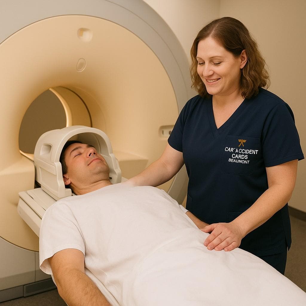
- Positioning: An MRI technologist will escort you to the MRI suite and help you lie down on a comfortable padded table. Coils specifically designed for the body part being imaged will be placed around or near the area.
- Entering the Scanner: The table will slide into the opening of the MRI machine. The body part being scanned will be positioned in the center of the magnetic field.
- The Scan: The technologist will go to the control room and begin the scan. The machine will make loud knocking, buzzing, and whirring noises as it acquires images. This is normal. You must remain as still as possible during the scan sequences, which may last from a few seconds to several minutes. You will be given earplugs or headphones to reduce the noise.
- Communication: You will have a way to communicate with the technologist during the scan, usually via an intercom system. You may also be given a call button to press if you need assistance.
- Duration: The length of the scan varies depending on the area being imaged and the number of sequences required, but typically ranges from 30 to 60 minutes.
Our technologists are experienced and trained to make your MRI scan Beaumont TX experience as comfortable as possible.
Post-Scan Procedures and Results Delivery
Once the scan is complete, the table will slide out of the machine, and you can get up.
- Immediate Aftermath: There are typically no restrictions after an MRI scan unless you received a sedative to help with claustrophobia, in which case you will need someone to drive you home. If you received contrast, you may be advised to drink extra fluids to help flush it out of your system.
- Radiologist Interpretation: The images acquired during your MRI scan Beaumont TX are sent to a radiologist – a medical doctor specializing in interpreting medical images. The radiologist will carefully review the images and generate a detailed report outlining their findings.
- Results Delivery: The time it takes to receive the report can vary, but at Car Accident Cares, we strive for efficiency, often providing same-day results when possible. The report will be sent to the doctor who ordered your MRI scan.
- Integration with Treatment: Your doctor will discuss the MRI findings with you and integrate them with your clinical symptoms and other diagnostic information to arrive at a diagnosis and develop or modify your treatment plan. Our team facilitates the seamless integration of your MRI scan Beaumont TX results with our treatment teams, including our pain management consultants, chiropractors, and other specialists, ensuring timely and coordinated care.
Unique Advantages of Our Beaumont MRI Services
Choosing Car Accident Cares for your MRI scan Beaumont TX offers distinct advantages, especially when you need rapid and affordable diagnostic imaging after an injury:
- 24/7 Availability: We understand that injuries don’t happen on a schedule. Our MRI scan Beaumont TX services are available 24 hours a day, 7 days a week, allowing you to get scanned promptly without long wait times often associated with hospital imaging departments.
- Affordable Pricing: We are committed to transparent and affordable pricing, often significantly lower than hospital rates. This is particularly helpful for patients managing healthcare costs after an accident. Explore our MRI cost solutions.
- Trauma-Trained Technologists: Our technologists are experienced in working with patients who have suffered injuries, providing compassionate and skilled care during the scan.
- Focus on Injury Diagnosis: Our services are specifically geared towards diagnosing injuries, ensuring our radiologists are experienced in interpreting scans related to trauma, including those from car accidents.
We aim to provide high-quality, accessible diagnostic imaging Beaumont to support your recovery journey.
What MRI Scans Don't Show
While MRI is incredibly powerful, it’s not the ideal imaging modality for every situation. Understanding its limitations helps manage expectations and ensures the most appropriate imaging test is selected for your specific needs.
Limitations in Acute Bleeding Detection
In emergency situations, particularly after severe trauma, a CT scan is typically the preferred initial imaging modality for evaluating internal injuries. This is because CT is significantly faster than MRI and is excellent at detecting acute bleeding fresh hemorrhage and bone fractures quickly. While MRI can detect some types of bleeding, especially chronic blood products, it is less sensitive and slower for identifying acute, active bleeding in trauma compared to CT. Therefore, in cases of suspected major internal bleeding, a CT scan may be performed first.
Challenges with Certain Metallic Objects
Although screening protocols are in place, certain types of metallic objects and medical implants can pose challenges or contraindications for MRI. As mentioned earlier, some pacemakers, older aneurysm clips, and certain cochlear implants are not MRI-compatible and mean a patient cannot safely undergo an MRI scan. Even MRI-safe or conditional implants can sometimes cause artifacts distortions on the images if they are located near the area being scanned, potentially obscuring visualization of nearby tissues. Thorough screening is always necessary.
Situations Requiring Alternative Imaging
There are scenarios where imaging modalities other than MRI might be a better initial choice or provide complementary information:
- Acute Bone Fractures: While MRI can detect occult fractures, X-rays and CT scans are generally faster and more readily available for initially evaluating suspected bone breaks.
- Evaluation of the Lungs or Chest: CT scans are typically better for visualizing the lungs and bony structures of the chest.
- Initial Evaluation of Major Trauma: As noted, CT is often the first-line imaging for widespread assessment in severe accidents due to its speed and ability to detect acute bleeding and fractures.
- Certain Vascular Issues: While MRA (MRI Angiography) exists, CT Angiography (CTA) may be preferred in some acute vascular emergencies.
- Dynamic Imaging: Ultrasound or fluoroscopy may be used to evaluate structures in motion, which MRI cannot do.
Often, different imaging modalities complement each other. An X-ray might show a possible issue, and an MRI scan Beaumont TX is then used to get a detailed look at the soft tissues involved. Learn when X-rays complement MRI findings or how CT scans are used by visiting our Best Affordable CT Scan Beaumont TX page. Choosing the right imaging test is a decision made by your doctor based on your symptoms and suspected injury.
Pediatric and Special Population MRI in Beaumont
Obtaining an MRI scan Beaumont TX can present unique considerations for certain patient populations, such as children, those with claustrophobia, or pregnant individuals, particularly in the context of trauma. Our facility aims to provide safe and accommodating services for these groups.
MRI Safety for Children After Accidents
Imaging children after an accident requires special care and expertise. While MRI is safe in terms of radiation, keeping a child still for the duration of the scan can be challenging.
- Sedation Protocols: For very young children or those who are unable to remain still, sedation may be necessary to obtain diagnostic quality images. This is done under the supervision of trained medical professionals.
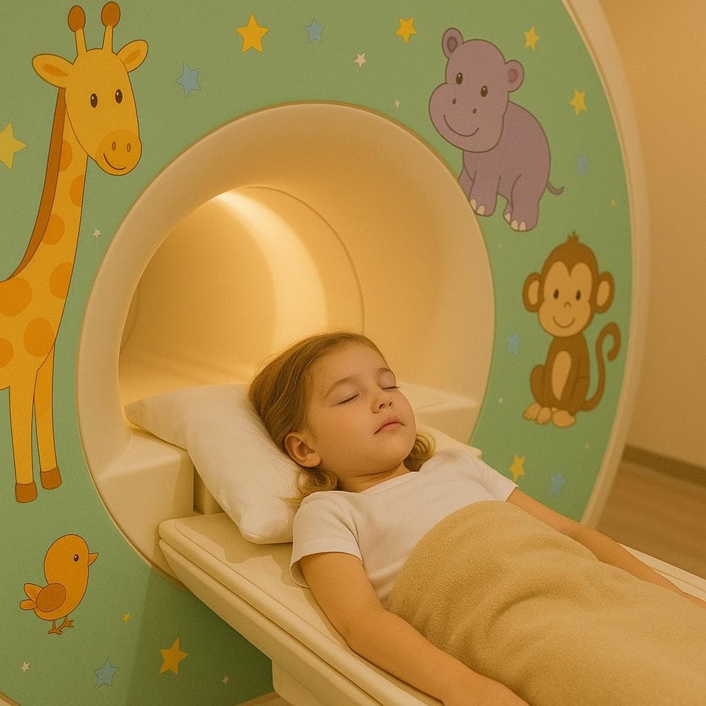
- Child-Friendly Techniques: Technologists experienced in pediatric imaging can use techniques to help children feel more comfortable, such as explaining the process in simple terms, allowing a parent to be present in the scanning room after safety screening , and using headphones that play music.
Ensuring a calm and safe environment for pediatric MRI scan Beaumont TX is a priority.
Accommodations for Claustrophobic Patients
Claustrophobia is a common concern with traditional closed MRI scanners. At Car Accident Cares in Beaumont, we understand this and offer strategies to help:
- Open MRI Options: If available, an open MRI system can provide a less enclosed scanning experience, which is often more comfortable for individuals with claustrophobia or those who are larger in size.
- Anxiety Management Strategies: Techniques like deep breathing exercises, guided imagery, or listening to music through headphones can help reduce anxiety during the scan.
- Sedation: In some cases, a mild sedative may be prescribed by your doctor to help you relax during the procedure.
Discuss your concerns about claustrophobia with your doctor or our staff when scheduling your MRI scan Beaumont TX.
Pregnancy Considerations in Trauma Imaging
When diagnostic imaging is needed during pregnancy, particularly after trauma, the safety of both the mother and the fetus is paramount. MRI is generally considered safer than X-rays or CT scans during pregnancy due to the absence of ionizing radiation.
- ACR Guidelines: The American College of Radiology (ACR) guidelines provide recommendations for imaging pregnant patients. MRI is often the preferred cross-sectional imaging modality when ultrasound is insufficient and radiation should be avoided.
- Contrast Use: Gadolinium-based contrast agents are generally avoided during the first trimester of pregnancy and used cautiously in the later trimesters, only when deemed medically necessary and the potential benefits outweigh the risks.
- 1st Trimester Precautions: While MRI is considered relatively safe throughout pregnancy, some practitioners may prefer to avoid non-urgent MRI in the first trimester as a precaution.
Decisions about imaging during pregnancy, including an MRI scan Beaumont TX after an accident, are made on a case-by-case basis in consultation with the patient’s healthcare providers, weighing the urgency of the clinical question against any potential risks.
Interpreting Your MRI Results
Once your MRI scan Beaumont TX is complete and interpreted by a radiologist, understanding the findings is the next crucial step in your recovery journey. The radiology report contains technical terms, and correlating the imaging findings with your symptoms requires clinical expertise.
Reading the Radiology Report
The radiology report is a written document detailing the findings of the MRI scan. It is generated by the radiologist who reviewed your images. The report will describe the appearance of the organs and tissues in the scanned area and note any abnormalities. Common terms you might see include:
- “Herniation” or “Bulge”: Refers to an intervertebral disc that is extending beyond its normal boundaries, potentially pressing on nerves.
- “Degenerative Changes”: Indicates age-related wear and tear in the spine, such as disc space narrowing or facet joint arthritis.
- “Signal Intensity”: Describes how bright or dark different tissues appear on the images, which can indicate inflammation, fluid, or other tissue characteristics.
- “Enhancement”: If contrast was used, this refers to areas that light up on the images after the contrast agent is injected, often indicating inflammation, infection, or tumors.
- “Stenosis”: Refers to a narrowing of a space, such as the spinal canal or the openings where nerves exit the spine.
Correlation with Clinical Symptoms
It’s important to understand that imaging findings on an MRI scan Beaumont TX do not always perfectly correlate with the level of pain or symptoms you are experiencing. For example:
- Asymptomatic Findings: It’s common for MRI scans, particularly in older individuals, to show disc bulges or degenerative changes that are not causing any pain or symptoms. These are considered incidental findings.
- Severe Pain, Mild Findings: Conversely, some individuals may experience significant pain even if the MRI shows only mild changes. This highlights the complex nature of pain, which can be influenced by inflammation, nerve sensitivity, and other factors not always fully captured by imaging.
Your doctor will take the information from your MRI scan Beaumont TX report and integrate it with your medical history, physical examination findings, and symptoms to arrive at an accurate diagnosis and develop a targeted treatment plan.
Second Opinion Opportunities
If you have questions or concerns about your MRI scan Beaumont TX findings or diagnosis, seeking a second opinion can provide additional perspective and peace of mind. At Car Accident Cares, we offer access to specialists who can review your images and report.
Consulting with our MD Consultation in Beaumont, TX service allows you to discuss your MRI results with experienced physicians who can provide further interpretation and discuss treatment options.
Cost and Insurance Considerations in Beaumont
Understanding the cost of an MRI scan Beaumont TX and how it relates to insurance coverage is a significant factor for patients, especially those recovering from an accident. We believe in transparency regarding pricing and assisting you in navigating insurance claims.
Understanding MRI Pricing Factors
The cost of an MRI scan can vary based on several factors:
- Facility Type: Hospital-based MRI centers often have higher overheads and therefore higher prices compared to independent imaging centers like Car Accident Cares.
- Body Part Scanned: The price may differ depending on the complexity of the area being imaged e.g., spine, brain, knee.
- Use of Contrast: Scans requiring a contrast agent are typically more expensive due to the cost of the agent and the additional time/expertise required for administration.
- Geographic Location: Healthcare costs can vary by region. Our facility in Beaumont is committed to providing affordable options within the local market.
- Equipment Type: Scanners with higher magnetic field strengths (e.g., 3T) or specialized capabilities may have different pricing.
Understanding these factors helps explain variations in diagnostic imaging Beaumont costs.
Navigating Texas Auto Insurance Claims
For individuals injured in car accidents in Texas, understanding how auto insurance can cover an MRI scan Beaumont TX is crucial.
- Personal Injury Protection (PIP): Texas law requires all auto insurance policies to include PIP coverage. PIP is no-fault coverage that pays for medical expenses including MRI scans, lost wages, and other related costs up to the policy limit, regardless of who was at fault for the accident. This is often the primary source of payment for medical bills after a crash.
- MedPay (Medical Payments Coverage): Similar to PIP, MedPay is another optional coverage that pays for medical expenses resulting from a car accident, regardless of fault.
- Liability Coverage: If the other driver was at fault, their bodily injury liability coverage may eventually pay for your medical bills, but this can take time to process.
- Letters of Protection (LOP) / Lien Agreements: In some cases, if you do not have sufficient PIP or MedPay, a diagnostic imaging Beaumont facility like Car Accident Cares may work with your attorney to provide services under a Letter of Protection or lien agreement, where payment is deferred until your case is settled.
Our team is experienced in working with auto insurance claims and can help you understand your coverage for an MRI scan Beaumont TX.
Self-Pay Options and Payment Plans
We are committed to ensuring that financial concerns do not prevent you from getting the necessary diagnostic imaging Beaumont. For patients who do not have insurance coverage or prefer to self-pay, we offer:
- Transparent Pricing: We provide clear, upfront information about the cost of your MRI scan Beaumont TX.
- Affordable Rates: Our self-pay rates are often significantly below typical hospital charges.
- Payment Plans: In some cases, we may be able to arrange a payment plan to spread the cost over time.
We encourage you to discuss your financial situation with our staff. We are here to help you explore options and access the MRI scan Beaumont TX you need. Contact our insurance specialists for assistance or to discuss payment options.
Future of MRI Technology in Beaumont
The field of MRI technology is continuously evolving, with advancements aimed at improving image quality, speed, and diagnostic capabilities. These innovations will continue to enhance the role of MRI scan Beaumont TX in diagnosing injuries and conditions.
3T MRI Advancements
Higher magnetic field strength scanners, such as 3T MRI, are becoming more common. A 3T MRI provides stronger signals than a 1.5T scanner, resulting in:
- Higher Resolution Images: This allows for the visualization of finer anatomical detail, which can be crucial for detecting subtle injuries like small ligament tears or cartilage damage that might be missed on lower field strength scanners.
- Faster Scan Times: In some cases, the stronger signal allows for shorter scan times while maintaining image quality.
These advancements in technology contribute to more precise diagnostic imaging Beaumont.
Functional MRI (fMRI) for Pain Mapping
Functional MRI (fMRI) is a specialized type of MRI that measures brain activity by detecting changes in blood flow. While primarily a research tool currently, fMRI holds promise for helping to understand and potentially map how the brain processes chronic pain. This could potentially lead to more targeted pain management for back pain strategies in the future. While not typically used for standard injury diagnosis, the potential applications of advanced MRI techniques in understanding chronic pain are an exciting area of development.
AI-Assisted Injury Detection
Artificial intelligence and machine learning algorithms are increasingly being applied to medical imaging. AI tools are being developed to assist radiologists in interpreting MRI scans, potentially:
- Reducing Interpretation Time: AI can quickly analyze images and highlight potential areas of concern.
- Improving Detection of Subtle Findings: Algorithms trained on large datasets may be able to identify subtle abnormalities that could be overlooked by the human eye.
- Quantifying Findings: AI can help provide objective measurements of injury severity or degenerative changes.
While AI is a tool to assist, the final interpretation of your MRI scan Beaumont TX will always be made by a qualified radiologist. These technological advancements underscore the commitment to providing cutting-edge diagnostic imaging Beaumont.
Conclusion
Understanding the capabilities of an MRI scan Beaumont TX is vital for anyone dealing with the aftermath of an accident or struggling with persistent pain. As a powerful, non-invasive diagnostic imaging Beaumont tool, MRI provides unparalleled detail of soft tissues, making it indispensable for diagnosing a wide range of injuries and conditions, from whiplash and herniated discs to traumatic brain injuries and hidden fractures.
At Car Accident Cares in Beaumont, we are dedicated to providing accessible, affordable, and high-quality MRI services to the community. Our 24/7 availability ensures you can get the crucial imaging you need when you need it, without unnecessary delays. Combined with our transparent pricing and experienced team, we aim to make the diagnostic process as smooth and informative as possible, setting the stage for effective treatment and recovery.
Don’t let uncertainty about your injuries hold back your recovery. Getting a detailed MRI scan Beaumont TX is a critical step towards understanding what’s happening internally so you can receive the most appropriate care.
To explore comprehensive imaging options available at our facility, please visit our page dedicated to Diagnostic Imaging. To find our location and get directions for your scan, visit our Find Us page.
Medically reviewed by
Verified
DS
Deepak Sharma
DC
Owner & Doctor of Chiropractic
Deepak Sharma, DC is a licensed Doctor of Chiropractic and the owner of Car Accident Cares in Beaumont, TX. He reviews the clinical content published across the site and works with a multidisciplinary network of MDs, chiropractors, imaging centers, and pain-management specialists focused on accurate diagnosis, evidence-based treatment, and complete recovery for motor-vehicle-accident victims across Beaumont and Houston.