Understanding MRI Scans: What They Show – Comprehensive Guide for Beaumont Patients
DS
By Deepak Sharma, DC
Medically reviewed
Owner & Doctor of Chiropractic · 9 min read · Published · Updated · Last reviewed
Introduction
After an accident, or when dealing with persistent pain, you might find yourself facing a swirl of medical terms and diagnostic uncertainties. One of the most powerful tools in modern medicine for uncovering hidden injuries is the MRI scan. With approximately 40 million MRI scans performed annually in the United States, this diagnostic tool has become essential for accurate injury assessment. If you’re in Beaumont, TX, and your doctor has recommended an “MRI scan Beaumont TX,” you might have questions about what it involves and what it can reveal.
Magnetic Resonance Imaging (MRI) is an advanced form of diagnostic imaging services that provides incredibly detailed pictures of soft tissues, organs, bone, and virtually all internal body structures. The global MRI market is projected to reach $12.51 billion by 2033, reflecting the growing importance of this technology in healthcare. This guide is designed to demystify the MRI process for Beaumont residents. We’ll explain how this cutting-edge technology works, what you can expect during the experience, and crucially, the types of injuries and conditions it is uniquely suited to diagnose, helping you move closer to effective treatment and recovery.
What is an MRI? Breaking Down the Technology
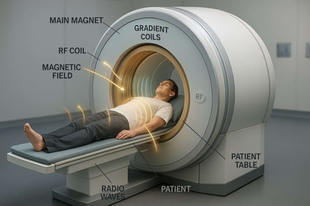
At its core, “what is an MRI”? MRI stands for Magnetic Resonance Imaging, a powerful non-invasive medical imaging technique. Unlike X-rays or CT scans, MRI does not use ionizing radiation. Instead, it employs a strong magnetic field and radio waves to create detailed cross-sectional images of organs and soft tissues within the body. MRI demonstrates the highest level of efficacy in injury identification, detecting 85% of injuries with 95% specificity, making it superior to other imaging modalities.
The technology behind magnetic resonance imaging works by temporarily realigning the hydrogen atoms that are abundant in water molecules within your body. When the radiofrequency current is turned off, these hydrogen atoms return to their original alignment, releasing energy. The MRI scanner detects this released energy, and a computer then processes the signals to create highly detailed images. This makes MRI exceptionally good at distinguishing between different types of soft tissues, which can be difficult to visualize with other methods. To learn more about our best affordable MRI diagnostic imaging service, feel free to explore our site.
The MRI Process: What to Expect
Understanding the MRI process can ease any anxieties you might have. Knowing what to expect before, during, and after your scan helps ensure a smooth experience. Studies show that MRI scans in the U.S. are expected to grow by 18.3% by 2030 to reach a total scan volume of 33.1 million, indicating the increasing reliance on this diagnostic tool.
Preparation Guidelines
Before your MRI, you’ll receive specific instructions. It’s crucial to inform your technician about any metal in or on your body, including pacemakers, cochlear implants, certain tattoos, and metal fragments from previous injuries. You’ll typically be asked to change into a gown and remove all metal objects like jewelry, watches, and hairpins. In some cases, a contrast dye may be administered intravenously to enhance the visibility of certain structures. If you experience claustrophobia, discuss this with your doctor beforehand, as options like open MRI machines or mild sedatives might be available. Most MRI scans take between 30 to 60 minutes, though some can be longer depending on the area being scanned.
During the Scan
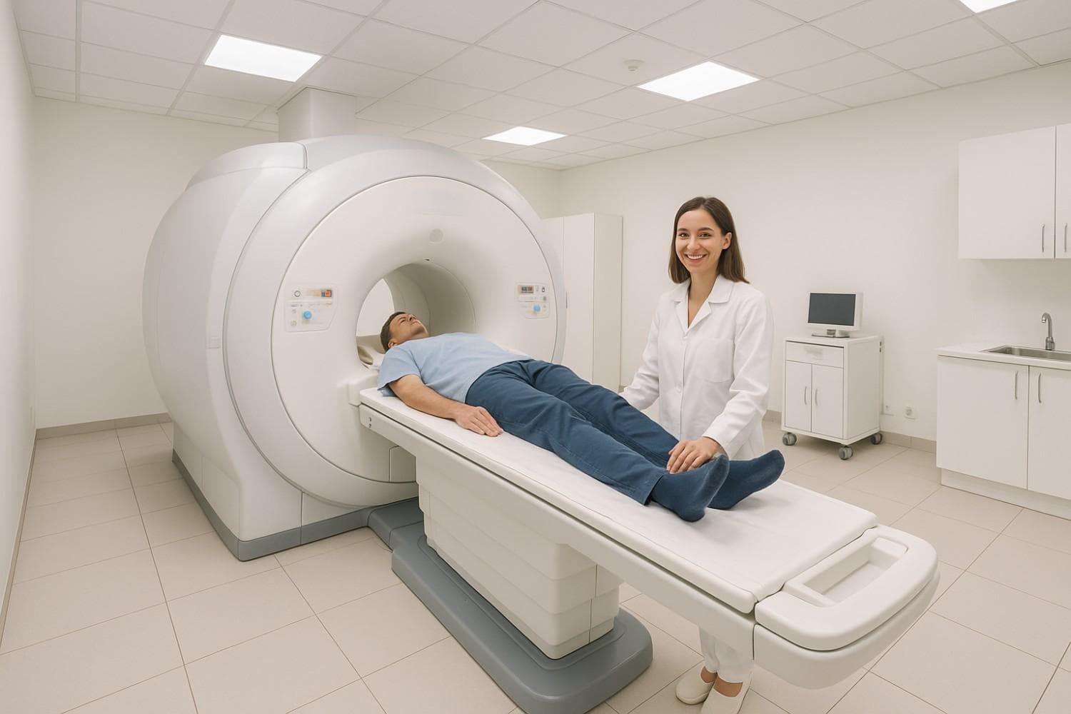
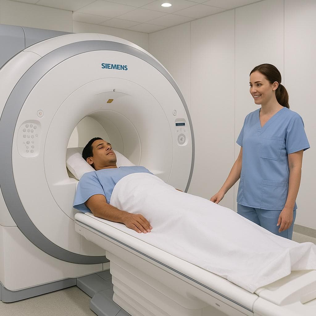
During the scan, you’ll lie on a movable table that slides into the MRI machine, which resembles a large, tube-like tunnel. The machine produces loud knocking or thumping sounds as it works, so you’ll be given earplugs or headphones to wear. It’s vital to remain as still as possible to ensure clear images. You’ll be able to communicate with the technician via an intercom system throughout the procedure, and they will be monitoring you closely from an adjacent room.
Post-Scan Process
Once your scan is complete, you can typically resume your normal activities immediately, unless you received sedation. The images will then be interpreted by a radiologist, and the results are usually available within 24-48 hours. Your referring doctor will then discuss the findings with you. In some cases, follow-up tests or additional imaging may be recommended if the MRI reveals complex findings. For urgent imaging needs or to discuss your results, please contact our office for timely assessment.
Key Injuries and Conditions MRI Detects
MRI’s unparalleled ability to visualize soft tissues makes it indispensable for diagnosing a wide range of injuries and conditions, particularly after accidents. Research demonstrates that MRI has a sensitivity of 93% for detecting spinal cord injuries and 94.4% for posterior longitudinal ligament injuries, making it highly effective for accident-related trauma assessment.
Soft Tissue Damage
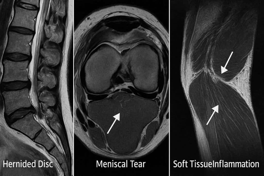
MRI is the gold standard for detecting damage to ligaments, tendons, and muscles. This includes:
- Ligament Tears: Such as ACL tears in the knee or rotator cuff tears in the shoulder. Studies show MRI has a sensitivity of 100% for ACL injury detection with 96.6% specificity.
- Muscle Strains: Identifying the extent and location of muscle tears.
- Tendon Injuries: Diagnosing conditions like Achilles tendon ruptures or tendinitis.
This is especially crucial in car accident contexts, where injuries like whiplash neck pain treatment often involve soft tissue injuries. With approximately 3 million Americans sustaining whiplash injuries annually, MRI becomes essential for accurate diagnosis as these injuries often involve soft tissue damage that X-rays cannot fully show.
Spinal Injuries
For back and neck pain, MRI provides detailed views of the spine’s delicate structures:
- Disc Herniations: Clearly showing when a spinal disc has ruptured or bulged, potentially pressing on nerves. Research indicates that lumbar disc herniations occur in up to 35% of individuals who experience moderate to severe spinal trauma from motor vehicle accidents. Learn more about herniated disc doctor diagnosis.
- Spinal Cord Compression: Identifying any narrowing of the spinal canal that is compressing the spinal cord.
- Nerve Root Impingement: Pinpointing where nerves are being pinched or irritated, a common cause of sciatica and radiating pain.
These findings are vital for developing effective lower back knee pain treatment strategies and pain management clinic approaches.
Joint and Bone Abnormalities
Even though X-rays are primary for bones, MRI can offer deeper insights:
- Hairline Fractures: Detecting subtle bone fractures that may not be visible on X-rays.
- Cartilage Damage: Visualizing damage to the cartilage in joints, crucial for conditions like arthritis or sports injuries.
- Bone Tumors and Infections: Identifying abnormal growths or infections within the bone marrow.
These detailed images can guide specialists, including those who may recommend an orthopedic and spine surgeon consultation, if necessary.
Traumatic Brain Injury Detection
MRI is particularly effective for traumatic brain injury assessment following accidents. Motor vehicle collisions account for approximately 280,000 traumatic brain injuries annually in the United States, and TBIs from motor vehicle crashes result in 218,936 emergency department visits, 56,864 hospitalizations, and 16,402 deaths annually. MRI’s superior soft tissue visualization makes it invaluable for detecting:
- Diffuse Axonal Injury: Microscopic brain damage that CT scans often miss
- Subtle Brain Contusions: Bruising of brain tissue
- Microbleeds: Small hemorrhages that indicate brain trauma
White Matter Changes: Damage to brain’s communication pathways
Why Choose MRI Over Other Imaging Methods?
When it comes to understanding your injury, knowing which imaging method is best suited for your specific situation is key. While X-rays and CT scans have their place, MRI offers distinct advantages:
MRI vs. X-ray: X-rays are excellent for bones and fractures, but they don’t provide much detail for soft tissues. MRI excels here, offering high-resolution images of ligaments, tendons, muscles, and nerve tissue.
MRI vs. CT Scan: CT scans also provide detailed cross-sectional images, often faster than MRI and with good bone detail. However, CT scans use X-rays (ionizing radiation), while MRI is radiation-free. MRI is generally superior for visualizing soft tissue conditions and certain brain or spinal cord issues. For comprehensive imaging options, you can explore our best affordable CT scan Beaumont TX services.
MRI vs. Ultrasound: Ultrasound uses sound waves and is good for real-time imaging of superficial soft tissues and blood flow, but it lacks the depth penetration and comprehensive view of an MRI for deeper structures.
Each modality has its best use. For alternative imaging options, you can discuss with our X-ray Beaumont TX experts. The choice depends on the suspected injury and the specific information needed for diagnosis. If you’re unsure which imaging is right for you, or to discuss your imaging needs, we encourage you to discuss your imaging needs with our specialists.
Answer Engine Optimization: Common Questions About MRI
What does an MRI show that other tests cannot? MRI provides superior soft tissue visualization, detecting injuries to ligaments, tendons, muscles, and nerve tissue with 85% sensitivity and 95% specificity. Unlike X-rays or CT scans, MRI can identify subtle brain injuries, spinal cord damage, and soft tissue trauma that other imaging methods might miss.
How accurate is MRI for detecting car accident injuries? MRI demonstrates exceptional accuracy for accident-related injuries, with 93% sensitivity for spinal cord injuries and 94.4% for ligament damage. For specific injuries like ACL tears, MRI achieves 100% sensitivity with 96.6% specificity, making it the gold standard for post-accident evaluation.
When should I get an MRI after a car accident? You should consider an MRI if you experience persistent pain, neurological symptoms, or suspected soft tissue injuries that X-rays cannot detect. With approximately 869,000 cervical spine injuries occurring annually from traffic crashes, early MRI evaluation is crucial for proper treatment planning.
Conclusion
An MRI scan is an invaluable diagnostic tool, particularly when assessing injuries sustained in accidents or when pinpointing the cause of persistent pain. For Beaumont residents, understanding what an MRI shows can provide peace of mind and pave the way for effective treatment. With the global MRI market growing at 6.52% annually and technology advancing rapidly with AI integration, MRI continues to evolve as a cornerstone of modern medical diagnosis.
MRI’s ability to provide detailed, radiation-free images of soft tissues, nerves, and subtle bone abnormalities makes it a crucial component of modern medical diagnosis. Given that motor vehicle collisions account for 37.21% of traumatic spinal cord injuries and approximately 20% of all TBIs annually, MRI’s role in accident-related injury assessment cannot be overstated.
Our team at Car Accident Cares is dedicated to providing accessible and comfortable MRI services, ensuring you receive the clearest possible picture of your condition. With advanced technology and expert interpretation, we help ensure accurate diagnoses, setting you on the right path to recovery. Don’t let diagnostic uncertainty prolong your discomfort.
Medically reviewed by
Verified
DS
Deepak Sharma
DC
Owner & Doctor of Chiropractic
Deepak Sharma, DC is a licensed Doctor of Chiropractic and the owner of Car Accident Cares in Beaumont, TX. He reviews the clinical content published across the site and works with a multidisciplinary network of MDs, chiropractors, imaging centers, and pain-management specialists focused on accurate diagnosis, evidence-based treatment, and complete recovery for motor-vehicle-accident victims across Beaumont and Houston.