The Difference Between a CT Scan and an MRI in Beaumont, TX
DS
By Deepak Sharma, DC
Medically reviewed
Owner & Doctor of Chiropractic · 6 min read · Published · Updated · Last reviewed
Introduction
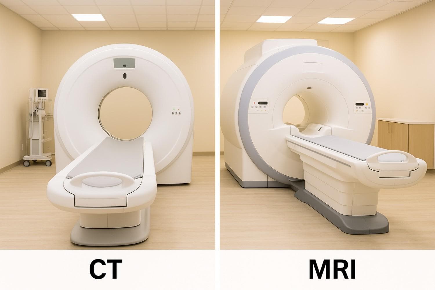
Diagnostic imaging plays a pivotal role in modern medicine, with Computed Tomography (CT) and Magnetic Resonance Imaging (MRI) among the most commonly ordered tests. For Beaumont, TX patients, understanding the difference between CT scan and MRI helps clarify why a physician might recommend one modality over another. While both generate cross-sectional images of internal anatomy, they leverage distinct technologies, excel at visualizing different tissues, and carry unique considerations in terms of speed, cost, and safety. This guide provides a comprehensive comparison—enabling Beaumont residents to make informed decisions about their medical imaging differences options.
Fundamental Technology Differences
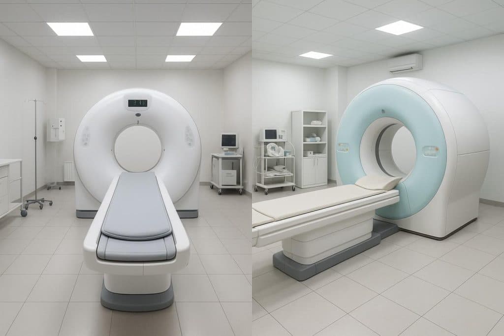
The core difference between CT scan and MRI lies in their underlying technology. While both provide detailed internal views of the body, they achieve this through entirely different means. Understanding these fundamental distinctions is crucial for grasping why each scan excels in different diagnostic situations.
How CT Scans Work
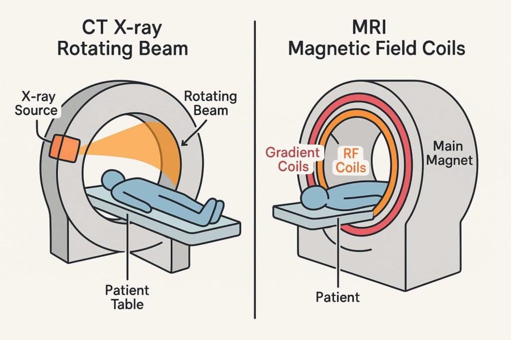
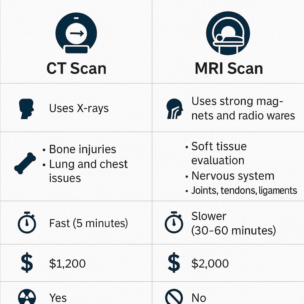
A CT scan uses diagnostic X-rays to capture multiple projections of the body as an X-ray tube rotates around the patient. Detectors opposite the source measure transmitted radiation, and advanced computer algorithms reconstruct slice-by-slice images of bones, organs, and vessels. These slices can be reformatted into 3D models, aiding in surgical planning and trauma assessment. A typical CT exam lasts 2–10 minutes, making it ideal for emergency settings.
How MRI Scans Work
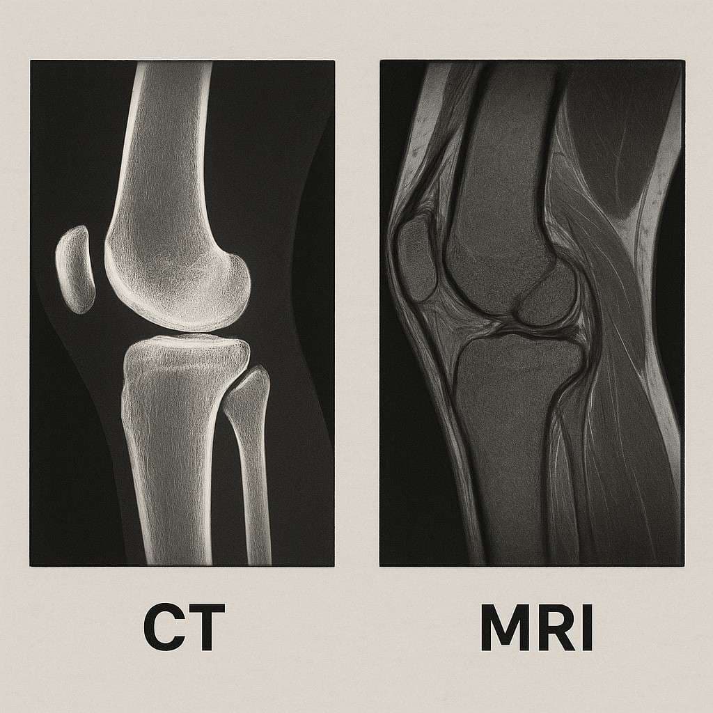
In contrast, MRI employs a strong magnetic field (commonly 1.5–3 Tesla) and radiofrequency pulses to align and then perturb hydrogen protons in water molecules. As protons realign, they emit signals captured by receiver coils and processed into high-resolution images, particularly of soft tissues. MRI exams generally take 15–45 minutes, depending on the number of sequences and use of contrast agents.
Diagnostic Strengths and Clinical Applications
CT and MRI are complementary technologies, each with distinct diagnostic strengths based on the types of tissues they visualize best and the medical conditions they are designed to assess. Knowing these strengths helps clarify the difference between CT scan and MRI.
CT Scan Imaging Strengths
CT scans provide unparalleled visualization of dense structures and acute pathology, including:
- Bone Detail: Detects fractures, joint injuries, and spinal anatomy with high accuracy.
- Emergency Trauma: Rapid identification of intracranial hemorrhage, visceral organ injury, and vascular compromise in trauma patients.
- Lung Imaging: Excellent for pneumonia, pulmonary embolism, and lung nodule detection.
- Calcifications and Stones: Clear depiction of vascular calcifications and renal or biliary stones.
- Cancer Staging: Efficiently evaluates chest, abdomen, and pelvis for tumor extent.
MRI Imaging Advantages
MRI excels at soft tissue contrast without ionizing radiation, making it the go-to study for:
- Neurological Conditions: Superior for brain tumors, multiple sclerosis plaques, and spinal cord lesions.
- Musculoskeletal Injuries: Detailed assessment of ligaments, tendons, cartilage, and bone marrow edema in sports injuries.
- Abdominal and Pelvic Organs: High sensitivity for liver lesions, pancreatic tumors, and gynecologic pathology.
- Certain Cancers: Optimal for prostate, breast, and select brain tumor evaluations.
Speed, Cost, and Accessibility
Beyond technology, practical considerations like time, cost, and accessibility also play a role in the difference between CT scan and MRI and which is chosen.
Time Requirements and Emergency Use
CT’s swift acquisition 2–10 minutes renders it indispensable in acute scenarios such as suspected stroke, trauma, or pulmonary embolism. MRI’s longer duration (15–45 minutes) suits non-urgent studies where maximal tissue characterization is prioritized.
Cost Considerations
CT exams typically range from $500 to $3,000, while MRI costs average $1,200 to $4,000, depending on anatomic region and use of contrast. Both modalities often require prior authorization. Discuss financial options with your insurer and our billing office to manage out-of-pocket expenses.
Accessibility and Patient Factors
CT scanners accommodate most patients, including those with pacemakers or metal implants, due to minimal interference with X-ray. MRI imposes strict metal screening protocols because strong magnetic fields can interact with ferromagnetic objects. Claustrophobic individuals may need anxiolytics or open-bore MRI systems.
Safety Considerations
Patient safety is paramount. Understanding the safety profiles is another key difference between CT scan and MRI.
CT Radiation Exposure
CT uses ionizing radiation, with effective doses ranging from 1–15 mSv depending on the body region. While the risk of radiation-induced malignancy from a single CT is very low, cumulative exposure is monitored, particularly in pediatric patients.
MRI Safety Profile
MRI involves no ionizing radiation. Primary safety concerns include interactions with metallic implants or devices and the possibility of heating. Thorough pre-scan screening prevents complications. Gadolinium-based contrast agents have a strong safety record, with adverse reaction rates under 0.5% and severe reactions in fewer than 0.01% of administrations.
Clinical Decision Making: When to Choose CT vs MRI
Understanding when to get CT vs MRI is about recognizing the specific clinical situations where each excels. Doctors base their choice on the patient’s symptoms, medical history, and the specific diagnostic question they need to answer.
Conditions Best Suited for CT
Physicians often select CT for:
- Trauma Assessment: Rapid detection of hemorrhage and organ injury.
- Bone Fractures: Complex fracture mapping, especially of the spine and pelvis.
- Pulmonary Evaluations: Pneumonia, embolism, or mass detection.
- Abdominal Emergencies: Appendicitis, bowel obstruction, or kidney stones.
- Post-Surgical Hardware: Imaging around metal implants with minimal artifact.
Conditions Best Suited for MRI
MRI is preferred for:
- Soft Tissue Evaluation: Ligament tears, muscle strains, and cartilage damage.
- Spinal Pathology: Herniated discs, spinal stenosis, and nerve compression.
- Neurological Disorders: Stroke, brain tumors, and demyelinating diseases.
- Cancer Characterization: Prostate, breast, and select liver tumors.
Choosing the Right Imaging Center in Beaumont
Selecting an accredited facility with board-certified radiologists and subspecialty expertise ensures diagnostic accuracy. Beaumont centers offering integrated care coordinate seamlessly with treatment teams, expediting follow-up and interventions. Locate our accredited Beaumont imaging services for scheduling and preparation details.
Future Developments in CT and MRI
Advancements such as dual-energy CT, ultra-high-field (3T and 7T) MRI, functional MRI (fMRI), and diffusion tensor imaging continue to expand diagnostic capabilities. These specialized applications guide surgical planning, radiation therapy, and interventional procedures, further blurring the lines between diagnostic imaging and treatment.
Conclusion
Understanding the difference between CT scan and MRI is key to making informed healthcare decisions. The choice of when to get CT vs MRI ultimately depends on your unique clinical needs, the urgency of your condition, and patient-specific factors.
Both CT and MRI are indispensable, complementary tools in modern diagnostic pathways. CT’s speed and bone visualization empower acute care, while MRI’s soft tissue resolution enhances evaluation of chronic and complex conditions. Beaumont, TX patients benefit most by understanding these differences and partnering with imaging specialists to select the most appropriate study.
Contact our diagnostic imaging team today to schedule a consultation and take the next step toward accurate diagnosis and effective treatment.
Medically reviewed by
Verified
DS
Deepak Sharma
DC
Owner & Doctor of Chiropractic
Deepak Sharma, DC is a licensed Doctor of Chiropractic and the owner of Car Accident Cares in Beaumont, TX. He reviews the clinical content published across the site and works with a multidisciplinary network of MDs, chiropractors, imaging centers, and pain-management specialists focused on accurate diagnosis, evidence-based treatment, and complete recovery for motor-vehicle-accident victims across Beaumont and Houston.